Thursday, January 31, 2013

URINARY PROBLEMS CAUSED BY SPINAL CORD INJURY


Management of Urinary Problems Caused by Spinal Cord Injury


Stephen Burns, MD, Staff Physician, SCI Service, VA Puget Sound Health Care System, Associate Professor, Dept. of Rehabilitation Medicine, University of Washington

Table of Contents


Before 1940, most people with spinal cord injuries died from urinary tract infections in the first few months after injury. After the introduction of antibiotics in the 1940's, people started surviving longer, but renal complications continued to be a problem and kidney failure became the leading cause of death. With current management practices and periodic testing, things have improved greatly, and now fewer than 3% of people with SCI die from kidney failure.

How the urinary system works


drawing showing the parts of the urinary tract

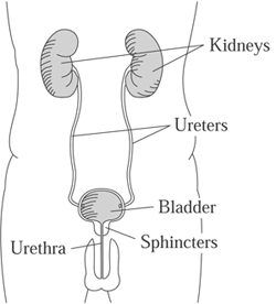
The upper urinary tract consists of the kidneys, which filter the blood and produce urine, and the ureters, which connect the kidneys to the bladder. The upper tract is not directly affected by spinal cord injury.

The lower urinary tract has muscles which are affected by the spinal cord injury: the bladder  muscle (detrusor) and the valve muscles (sphincters). The urethra connects to the outside of the body where the urine passes through.

In normal urination, the bladder is either filling or emptying. The bladder is relaxed as it fills with urine, and the sphincter stays closed during this time so the urine doesn’t leak out. When it’s time to empty, the bladder contracts and the sphincter relaxes so the urine can flow out. Most of the time the bladder is relaxed and filling as urine is made. The bladder and sphincter muscles are automatically coordinated to contract and relax at the correct time. These are reflex patterns wired into the brainstem and spinal cord.

After spinal cord injury, several different kinds of urinary problems can result, depending on the level of injury and which nerves and reflexes have been disrupted. Bladder and sphincter muscles may be weak, overactive or poorly coordinated.

Essentially, two basic problems occur. Filling problems (incontinence or leaking) occur when the bladder is overactive and contracts too much or at the wrong time, or the sphincter doesn’t contract enough to keep the urine from leaking out. Emptying problems (retention) occur when the bladder doesn’t contract enough or the sphincter won’t relax. Treatment will depend on what kind of problem you are having.


How we figure out what the bladder is doing


  • We can get a lot of the information just starting with the history of when and under what circumstances incontinence occurs.
  • A neurological exam can tell us what is happening with the whole nervous system in terms of strength, sensation and reflexes. This gives us a good picture of what is likely to be happening with the nerves to the bladder and sphincter, what is working and what's contracting and what's not.
  • A postvoid residual test shows how much urine remains in the bladder after voiding (emptying). This can be done using a catheter or by ultrasound.
  • A group of tests called urodynamics tells us more precisely what the nerves and bladder are doing during filling and emptying. These tests require the bladder to be slowly filled with fluid through a small catheter, while the activity of different muscles is measured.  Urodynamics help answer these types of questions:
    • Is the bladder relaxing enough to allow it to fill up with urine?
    • Is the sphincter opening at the right time?


Choosing the best method of bladder drainage


The goal in choosing a method of bladder drainage is to find the simplest, most convenient and least expensive method that will keep you dry, avoid serious complications and treatment side effects, and preserve your kidneys for your entire life. There are several different methods, depending on your injury and circumstances, and almost all of them give good outcomes, with just a few exceptions.

No catheter in the bladder


  • Voluntary voiding (bladder emptying) under normal control, possible in combination with medications to calm an overactive bladder muscle if necessary.
  • Involuntary voiding, where the bladder fills to a certain point, kicks off and empties. Emptying might occur spontaneously or in response to pressure on the bladder such as tapping the bladder (Crede) or bearing down (Valsalva).  These are NOT recommended for most patients, however, because they can cause problems such as high pressure that can damage the kidneys. For males, a condom catheter can be used to collect the urine.

Bladder emptying for males: Open the sphincter


There are a few methods for keeping the sphincter open so urine can flow freely from the bladder into a condom catheter.

  • Sphincterotomy: Surgically cut and open the sphincter. Scarring can occur over time, and the surgery may need to be repeated. It can also worsen erectile dysfunction.
  • Botox injected into the sphincter. This needs to be repeated every three to nine months, and as it wears off there is an increased chance for urinary retention.
  • Urethral stent (small steel tube) placed in the sphincter. Disadvantage are that the stent can move around or tissue may grow into it and block the flow or urine, requiring corrective surgery.

Methods that keep the sphincter open only work for people whose bladders are able to contract, allowing urine to continuously drain into a collection device like a condom catheter. If your bladder does not contract, the urine won’t drain out, and you are at risk for infection.

The downside of any sphincterotomy method is that the bladder may lose its ability to contract and urinary retention may develop over time. Also, condom catheters are not without problems. They can be hard to keep in place, and some patients will need to have a penile prosthesis put in so there is enough penis for the condom to attach to. And even though the condom catheter does not involve a tube going into the bladder, it does not seem to result in fewer UTIs than indwelling or Foley catheters.


Intermittent Catheterization (ICP)


This method is also known as ICP (Intermittent Catheterization Program), CIC (Clean Intermittent Catheterization) and I & O (In and Out) catheterization. With this method, you insert a catheter into the bladder and empty it completely every four to six hours. The goal is to cath frequently enough to keep urine volumes in the bladder lower than 500 ml. This method requires that you closely monitor your fluid intake, usually around 2 liters per day, otherwise you might be cathing too frequently to make this practical.

ICP is a preferred method for patients who have enough hand function (usually C7 and below, or C6 for motor incomplete injuries) to perform it independently and who can remember to cath on schedule. It is the closest thing to the normal bladder function, where the bladder fills continuously for a period of time and then empties all at once. This method reduces the risk for infections because there isn’t enough time for any bacteria left in the bladder to reproduce enough to cause symptoms.

Complications of ICP include narrowing of the urethra from passing the catheter through frequently. More rarely, inflammation of the epididymis (a duct that stores sperm) , hydronephrosis (enlargement of the urine collection section of the kidney) and reflux (backup of urine into kidney) may occur.

ICP is not usually a good method for someone who is unable to perform it independently. Having someone else cath you increases your risk for infections and also reduces your independence, since you need someone with you to perform the ICP.

Anticholinergic medications, such as oxybutynin (Ditropan) or tolterodine (Detrol), may be necessary to inhibit bladder contraction. Botox injection to the bladder muscle can also be used for this purpose.


Indwelling Catheter


An indwelling catheter is a common bladder-emptying method for those who cannot perform ICP. A tube is inserted into the bladder, where a balloon on the end holds it in place. It remains in the bladder and drains constantly into a container, such as a leg bag. There are two types of indwelling catheter:

  • Foley catheter: the tube is inserted through the urethra.
  • Suprapubic (SP): the tube goes through a hole in your abdomen.

Advantages:

  • It will usually empty the bladder and keep you dry regardless of what kind of bladder or sphincter problems you have.
  • Even those with higher level injuries can be completely independent—once you're set up, even if you have a high level injury, you can use an electric leg bag opener to empty out urine and not need assistance from anybody all day long.

Disadvantages:

  • Having a catheter sitting in the urethra all the time can cause urethral erosions, which is often a reason for switching to a suprapubic tube.
  • The suprapubic tube requires surgery, and sometimes the bladder neck needs to be closed to prevent leaking.
  • There is a catheter coming out of your body and a bag of urine with you all the time. Some people just don’t want that.
  • Increased risk of bladder cancer and bladder stones.
  • More infections than with ICP.


What’s different about females?


Because women have no penis, collecting urine is more difficult. There is no good external collection device, like a condom catheter, for women. Women doing ICP have more problems with incontinence than men because the female urethra is short and more likely to leak urine.

Women get different complications from having an indwelling Foley catheter for a long time. The urethra can become dilated (larger), which results in more leakage. Switching to a larger catheter just dilates the urethra more, causing more incontinence. For this reason, a suprapubic (SP) tube is a good option for a woman who otherwise would be using a Foley catheter.

What is the best method for you?


Considerations:

  • Do you have the hand function to do ICP independently?
  • How much mobility is required? For example, does the method require transferring to a toilet?
  • How much of the day is going to be devoted to bladder management?
  • What are the risks if you don’t follow the program? Are you likely to comply?
  • Do you live in a remote location with no follow-up around, or are you close to specialized medical care?
  • What's the likelihood that you would benefit from one of the more complicated, more time-intensive techniques?


 Other surgical options


  • An artificial urinary sphincter can be placed if there is incontinence due to the sphincter being open. This is a device that is surgically implanted in the body to substitute for the sphincter muscles. They have not been used commonly in people with SCI, since implanted devices are prone to infection, but some urologists do recommend them for certain individuals with SCI.
  • Bladder augmentation, which uses a piece of the bowel to enlarge the bladder, may be a good option for someone doing ICP whose bladder doesn’t hold enough urine in spite of medications.
  • Urinary diversion (diverting the urine away from the urethra)
    • Urostomy, which uses a piece of bowel to create a connecting tube from the bladder to the outside of the body (like a colostomy does for stool). Urine drains out and collects into a bag fastened to the opening (called a stoma). This is usually a fall-back method in cases where there have been major complications that cannot be treated with other methods.
    • Catheterizable stoma (Mitrofanoff) creates a thin tube from a piece of bowel that connects the bladder to the abdomen where a person can insert the catheter to drain the bladder (rather than inserting the catheter through the urethra).


Functional electrical stimulation (FES)


This system allows emptying without using a catheter. A surgically implanted stimulator and electrode trigger the bladder to squeeze when you flip a switch on an external stimulator. It requires cutting the sacral nerve roots, and you need to either use a condom catheter, a hand urinal, or transfer onto a toilet when the system is turned on. It can also be used to stimulate a bowel movement. It was on the market in the US (called the Vocare System) for a short time and continues to be available in Europe.

A new FES system currently under development at Case Western in Cleveland uses an electrode to block the sacral sensory roots so that you wouldn't need to cut the nerve roots. Somebody with an incomplete spinal cord injury could potentially use this method.


Botulinum toxin injection to the bladder


If oral medicines (anticholinergics) are unable to relax the bladder muscle enough for a person to do ICP, Botox injections to the bladder muscle can accomplish this. Botox is effective for about six to nine months. When it begins wearing off, you start having incontinence and need it done again.

Most common methods of urinary drainage five years after injury (SCI Model Systems data)


  Males Females
SP Tube 10% 7%
Foley 11% 23%
Condom Catheter 17% 0%
ICP 29% 26%
Normal 22% 28%


Urinary complications


Kidney and bladder stones


Stones are common in people with SCI. They can develop early on because large quantities of calcium leave the bones in the first few months after injury. It is more common to get stones later, and this is due to infections over the long term. Bacteria break down urea into chemicals that form stones, which can cause blockages, kidney damage and serious infections.

Hydronephrosis and reflux


These are similar conditions involving either a blockage of urine or a backwards flow of the urine up toward the kidney. It can have multiple causes, and the treatment is to remove whatever is blocking the system and to reduce the bladder pressure.

Bladder cancer


There is a small risk of bladder cancer for individuals using indwelling catheters. Screening recommendations are controversial since we don’t know who needs to be screened, how often, and how soon after injury. Unfortunately, these tend to be such aggressive cancers that even yearly screening won’t catch all of them because they grow so fast. Fortunately, bladder cancer is not very common.


Screening tests


We use a variety of tests to detect problems in the urinary system.

Lab tests


  1. Serum creatinine (blood test): Creatinine is filtered out by the kidneys. A high level in the blood means the kidneys are not filtering enough. To be useful, results must be monitored over time to see if there are changes. If it starts rising, it’s a sign something is wrong with the kidneys.
  2. Creatinine clearance: 24-hour urine collection to see how much filtering the kidneys are doing over time. This test may not give reliable results. Other lab tests are being studied as well to see what is best for screening.  

Imaging tests


  1. Ultrasound is a radiation-free, risk-free way to pick up on stones or blockages.
  2. CT scan of the kidneys, ureters and bladder (CT-KUB): uses lots of radiation and may carry a one in 3000 chance of producing a fatal cancer. While not recommended as a routine test, it is useful in specific situations.
  3. Renal scan: used to show kidney function, but image is fuzzy.

How often should the test be done?


Research has not established what testing should be done for everyone and how often. To some extent it should depend on the patient and what kinds of problems he or she is having. While early screening is not necessary for those who have fairly normal control of bladder, good sensation and not having symptoms, most people with spinal cord injury should have some sort of periodic testing of their urinary tract to detect problems before they become big problems.

The Consortium for Spinal Cord Medicine publishes a guideline for physicians stating that screening is usually done annually. However, since research has not established the necessary frequency for the screening tests, the guideline does not make a strong recommendation about how often the tests must be done.  (Bladder Management for Adults with Spinal Cord Injury: A Clinical Practice Guideline for Health-Care Professionals, www.pva.org). 


UTIs and antibiotics


When considering the use of antibiotics for UTIs, it is important to distinguish between actual infections and colonization.

  1. If you have bacteria in the urine (found through a lab test) AND have symptoms (fever, pain, spasticity), then you have an infection that needs to be treated with antibiotics.
  2. If you have bacteria in the urine but have NO symptoms, then you have what is called “colonization” and you should not be treated with antibiotics.

In general, treatment should be based on symptoms, rather than on bacterial count alone. Some bacteria don’t cause any symptoms, and their presence in the urine might even be keeping out other bacteria that could cause problems. In fact, there is currently some promising research into this idea of “bacterial interference” to determine whether inoculating people with a specific, relatively harmless bacteria will keep harmful bacteria away.

Prophylactic antibiotics, or taking antibiotics all the time to prevent UTIs, have not proven to be beneficial in research studies and can result in the proliferation of resistant bacteria that are hard to treat. A substance called methenamine, which turns into formaldehyde in the bladder, is used by some patients to try and reduce infections.

Cranberry (juice or tablets) has also been studied as a way of preventing UTIs. Usually the tablet form is used since drinking cranberry cocktail is so full of sugar and calories. Although cranberry has not proven effective in clinical trials with people who have SCI, it does seem to help some individuals. As with many aspects of bladder management after SCI, finding what works is often a matter of trial and error.


POSTED BY ATTORNEY RENE G. GARCIA:

For more information:- Some of our clients have suffered this kind of injuries due to a serious accident. The Garcia Law Firm, P.C. was able to successfully handle these types of cases. For a free consultation please call us at 1-866- SCAFFOLD or 212-725-1313.
http://sci.washington.edu/info/forums/reports/urinary_problems.asp

Monday, January 28, 2013

Post-Traumatic Stress Disorder


Definition

Post-Traumatic Stress Disorder (PTSD) is a specific form of anxietythat comes on after a stressful or frightening event.

Description

While some stressors are a predictable part of most people's lives - for example, bereavement or chronicillness - others lie outside the range of these common experiences. These include hurricanes, floods and other natural disasters; airplane accidents and car crashes where considerable injury occurs; rapeand assault; and armed combat, torture, and existence in a death camp. Such traumatic events would produce distress in almost everyone at any age. In some, they give rise to PTSD.

Causes

PTSD is one of the few psychological disorders with a clear point of origin. Although there are some people who have been exposed to trauma and do not develop PTSD, there are certain susceptibility characteristics in people who develop PTSD. These susceptibility factors include the following:

Previous chronic exposure to stress. Susceptibility depends primarily on the survivor's interpretation of the event. Thus, an event that is merely troublesome to one person can be devastating to another. Moreover, each of us may have a stress threshold, so that those who have endured more stress in life are considered more susceptible to PTSD.

Preexisting psychological disorders. People who suffer from depressionor anxiety or have a family history of anxiety are more likely to develop PTSD.

Inadequate support systems. A failure by loved ones to acknowledge the trauma, or loved ones who downplay its importance and place blame on the victim, increase the risk of PTSD.

Genetic predisposition.

Recent (acute) stressful life changes.

Feeling of lack of control. The state of having "learned helplessness" or giving total control to another individual can give rise to PTSD.

Recent excessive alcohol intake.


Symptoms

The post-traumatic reaction often begins immediately but may emerge after days, weeks, months or even years after the event. There are three kinds of symptoms, hyperalertness/hyperarousal, intrusion and emotional constriction or numbing.

1. Hyperalertness/hyperarousal.People are often edgy, irritable, easily startled, constantly on guard, have poor sleep habits, are agitated and find it difficult to concentrate.

2. Intrusion. People tend to re-experience the event in painful memories, flashbacks, dreams or nightmares. People with PTSD also may experience a worsening of their symptoms when they find themselves in situations that resemble the original trauma or that may be taken as symbols of it - for example, a spell of hot, humid weather for a Vietnam veteran, or a dark street for a woman who has been raped. In avoiding, or attempting to avoid, such situations, an individual's ability to function socially and at work may be severely impaired.

3. Emotional constriction or numbing. Another symptom is called "psychic numbing" or "emotional anesthesia." Sufferers exhibit a pervasive feeling of being detached from other people, from the outside world, and from activities that used to be enjoyable.

They also have a greatly diminished ability to experience emotion, especially tenderness and the feelings associated with intimacy and sex. Other symptoms reflect an over-aroused autonomic nervous system. Sufferers have difficulty falling asleep or staying asleep; they are keyed up and their startle response is heightened.

Anxiety and depression are common in those with PTSD. Irritabilityis a further problem. Guilt about surviving when others did not, and about the behavior that was necessary for survival, may be constant and painful. Some of those with this disorder turn to alcohol or drugs for escape, others may become self-defeating or suicidal.

Diagnosis

According the Diagnostic and Statistical Manual of Mental Disorders the following symptoms must be present for a diagnosis of PTSD:

  • Recurrent and intrusive distressing recollections or dreams about the event
  • Feeling as if the traumatic event were recurring, such as through hallucinations and flashbacks
  • Intense psychological distress at cues that symbolize the event

Additionally, these symptoms must endure for more than a month and must cause clinically significant distress or functional impairment.


Treatment

As with many other psychiatric disorders, the most effective treatments are psychotherapy and drug treatment.

Psychotherapy

If functional impairment occurs, or if the symptoms are severe and prolonged, some form of psychotherapeutic treatment is indicated.

With psychodynamic psychotherapy, the individual has the opportunity to discuss the event, relive it, and achieve a better understanding of why it has triggered so severe a psychological and physiological reaction. Cognitive-behavioral techniques may be used to reduce anxiety and depression, aid with insomnia, and treat any phobic symptoms that may have developed.

Psychotherapists treating PTSD try to provide a way for the patient to feel safe in confronting the traumatic event and linking it emotionally as well as intellectually to the symptoms. Patients must learn to think about the trauma without intrusive reliving and exercise self-control without avoidance and emotional numbing.

Some patients are reluctant to talk because they believe the therapist cannot possibly understand what they have been through. Some resist any suggestion that the traumatic event is related to their symptoms. Their helplessness is confirmed by nightmaresand flashbacks that remind them of their inability to protect themselves during the trauma. Some have become convinced that the world is hopelessly unjust, others that they themselves are defective or evil. Some patients make emotional demands and direct their accumulated anger at the therapist, they may quit therapy because they can no longer tolerate being exposed to their feelings about the trauma.

Drug Treatment

Drug treatment is aimed at relieving fear and anxiety and restoring normal sleep patterns. Tricyclic antidepressants, such as Tofranil and Elavil, or SSRIs such as Prozac or Zoloft, are helpful. Additionally, Catapres and Inderal are known to relieve agitation, while Xanax may help some people sleep.

POSTED BY ATTORNEY RENE G. GARCIA:


For more information:- Some of our clients have suffered this kind of injuries due to a serious accident. The Garcia Law Firm, P.C. was able to successfully handle these types of cases. For a free consultation please call us at 1-866- SCAFFOLD or 212-725-1313.

Thursday, January 24, 2013

TOTAL HIP REPLACEMENT: AN OVERVIEW

Introduction

Conventional, primary total hip replacement is a durable operation in the majority of patients. A hip replacement is a mechanical device with parts that are assembled before and during the operation, most commonly referred to as a “ball and socket.” After surgery, the prosthetic ball and socket restore movement in the hip during the life-span of the prosthesis (Figure 1).
Figure 1: An x-ray of a total hip replacement prosthesis with labels showing the "ball and socket."
As with any other mechanical device, a total hip replacement can be subject to various forms of mechanical or biological failure. Some of these conditions may require a re-operation of the hip replacement to address the cause of failure and its consequences. A re-operation of a total hip replacement is called a “revision.”

Why consider a hip revision?

The majority of elderly patients who receive a hip replacement retain the prosthesis for 15 to 20 years, and sometimes for life. However, some patients may need one or more revisions of a hip replacement, particularly if the initial hip replacement surgery is performed at a young age and the patient chooses to have a very active physical lifestyle.
Pre-operative investigations in patients who will undergo revision surgery are more extensive than those required in patients scheduled for a primary surgery. Often times, special radiographic (x-ray) projections, CT scan, or MRI of the hip may be necessary to determine position and fixation of the replacement parts (components), and to determine with precision the extent of bone loss around the failed implant. A preoperative aspiration (fluid sample) and/or special blood work may be needed if the surgeon suspects an infection in the failed hip.

Reasons for hip revision

Revision hip replacement is a relatively infrequent operation. In the United States, there are approximately 18 revision hip replacements performed for every 100 hip replacements.(1) The most frequent reasons for revision are:
  1. repetitive dislocation of a hip replacement
  2. mechanical failure (implant wear and tear – loosening - breakage)
  3. infection
Repetitive (recurrent) hip dislocation: As mentioned above, a hip replacement has a structure that resembles that of a natural hip (a ball and socket). For a hip replacement to function well, the ball has to be inside the socket at all times. The factors keeping the ball inside the socket are the alignment and fit of the ball and the socket, and the forces generated by the strong muscles and ligaments around the hip joint. A hip replacement is designed to have a large range of motion. However, some hip positions or trauma can force the hip ball out of the socket, a condition referred to as dislocation.(Figure 2)
Figure 2: An x-ray of a dislocated total hip replacement prosthesis showing the ball forced out of the socket.
Dislocations are relatively infrequent, particularly in the healthy patient who carefully follows precautions given by the surgeon and physical therapist. However, some patients are predisposed to this complication: the elderly, debilitated patients, or those who get a hip replacement after a hip fracture or after multiple hip surgeries.
Patients who suffered a hip dislocation are predisposed to additional dislocations, as the dislocated ball disrupts the important muscles and ligaments around the hip. Multiple dislocations are infrequent, but in patients who have suffered multiple hip dislocations, the orthopaedic surgeon may recommend revision surgery.
Revision surgery is effective in preventing a new dislocation. Prior to surgery, the surgeon may request imaging studies to determine the exact position and orientation of the different parts of the replacement. One or more parts may need to be re-oriented or completely exchanged during the revision.
In certain instances, the surgeon may use a device that “captures” the ball inside the socket (called a constrained socket). The proper healing of the soft tissues around a revised hip is most important for the success of the operation. Therefore, the surgeon may recommend wearing a brace for a few weeks after surgery. After surgery, it is important to follow the surgeon’s advice and to refrain from moving the hip into positions that can generate a new dislocation.
Mechanical wear, implant loosening, and breakage: The mobile parts of a hip replacement that move against each other will slowly wear and tear during the regular use of the replacement. The younger and the more physically active the patient is, the faster the wear. The wear consists of the generation of very small particles from constant, repetitive movement of the mechanic parts during the use of the hip. Depending on the type of hip replacement, the particles can be made out of plastic, cement, ceramic, or metal.
The patient’s immune system will recognize those particles as foreign and generate an immune response (like an allergic reaction). A strong reaction to the wear particles can result in the destruction of bone around the hip replacement (a condition called osteolysis). If the bone destruction is severe enough, the components of the replaced hip may become loose (Figure 3).
Figure 3: An x-ray of a loose total hip replacement prosthesis showing separation of the stem from the bone (as detailed by arrows).
A loose component can move against the surrounding bone, compounding the bone loss. If the bone loss is severe enough, a spontaneous bone fracture can occur (known as a pathologic fracture).
Mechanical wear and tear leading to loosening of the prosthesis (implant) is one of the most frequent forms of mechanical failure. However, other forms of mechanical failure are possible, like breakage of the prosthesis (Figure 4).
Figure 4: An x-ray detailing the broken stem section of a total hip replacement prosthesis.
During revision surgery for wear, mechanical loosening, or breakage, the surgeon will remove the worn/loosened/broken component/s, will assess the amount of bone loss, and will implant new components. Frequently, a bone graft (cadaveric bone from tested donors) is necessary to rebuild the bone content of the hip lost during the process of prosthetic failure. In certain instances, when there is a large amount of bone graft utilized, or when the patient’s bone stock is poor, the surgeon may ask the patient not to bear full weight in the operated leg for a period of time after surgery.
Infection of a hip replacement can occur at any time after surgery. The risk is higher in close proximity to surgery (first six weeks). However, after that, there is a low risk of “late” infections. Sometimes, infections in the mouth, gums, teeth, lungs, urine, or skin, or regular dental procedures that involve gum bleeding, can force bacteria in the blood stream. The bacteria can seed and infect a hip replacement, causing hip pain and fever.
In the presence of a prosthetic infection, the surgeon will make an attempt at identifying the organism (bacteria) that is causing the infection. A hip aspiration may be recommended. The liquid aspirated from the hip will be sent to the laboratory and tests performed to determine the type of bacteria present and the antibiotics the bacteria are sensitive to.
Once an infection in the hip replacement has been diagnosed, several treatment options are possible. The vast majority of treatments include surgery and a course of antibiotics specifically targeting the infecting bacteria. The treatment option depends on the type of bacteria, the sensitivity to antibiotics, the duration of the infection, the fixation of the hip replacement parts, and the patient’s general condition. The surgeon will discuss benefits and drawbacks of each treatment option.
The most common treatment options include:
  1. A thorough surgical cleaning of the hip replacement. This is generally recommended when the infection is discovered very early (within a few hours or days). Patients require six weeks of intravenous antibiotics and, frequently, a low dose of oral antibiotics for a long period of time (sometimes for life).
  2. A complete exchange of a hip replacement done in two stages: A first stage consists of the complete removal of the hip replacement, cleaning of the bone, and implantation of a temporary cement spacer that will allow some hip motion and deliver antibiotics to the hip area. This is generally followed by a six week course of intravenous antibiotics. The second stage consists of the re-implantation of a definitive hip replacement (generally 6 to 8 weeks after the initial operation).
  3. A complete exchange of a hip replacement done as a single operation, during which the infected prosthesis is removed, the bone is cleaned, and a new prosthesis is implanted. Patients require six weeks of intravenous antibiotics and, frequently, a low dose of oral antibiotics for a long period of time (sometimes for life).

Step-by-step animation of a single operation hip revision procedure


Preventing the need for revision hip surgery

The importance of awareness and postoperative surveillance: Some of the previously mentioned forms of failure can be prevented.
Dislocations can be prevented by following the surgeon’s instructions; some forms of hip infection can be prevented by prompt treatment of other bodily infections and by taking antibiotics before certain dental and other procedures.
The prosthesis’ natural wear and tear is generally painless. Therefore, it is very important that the patient has his/her hip replacement regularly checked. A simple physical examination and radiographs are necessary at the intervals designated by the surgeon. If excessive wear and/or bone loss is detected at any time, close monitoring is necessary to determine the best possible time (if any) to have the hip replacement revised.

Things to keep in mind when considering revision hip surgery

During revision surgery, the surgeon may need to remove or exchange one or more parts of the hip replacement. The parts that are not attached to the bone can be safely exchanged with minimal to no removal of the patient’s bone. However, if the metallic parts in contact with the bone need to be changed, some bone loss generally occurs. In addition, some of the musculature around the hip will be lost, thus affecting the strength of the hip and the patient’s function after surgery. The results of revision surgery are not as predictable as those of the primary surgery. Complications are more frequent.
A revision of a hip replacement is generally more challenging than the initial operation and it should not be regarded or compared to a “change of tires on the car.” The results of surgery and the durability of the revised hip replacement are less predictable than those of the primary operation. With every revision surgery, there is some loss of muscular mass, bone, or both. The duration of surgery is generally longer and the likelihood of complications higher than during the primary surgery.

Reference

1. Memtsoudis SG, Besculides MC, Gaber L, Liu S, González Della Valle A.Risk factors for pulmonary embolism after hip and knee arthroplasty: a population-based study. Int Orthop. 2009 Dec;33(6):1739-45.

 
POSTED BY ATTORNEY RENE G. GARCIA:


For more information:- Some of our clients have suffered injuries that require a Hip Replacement due to a serious accident. The Garcia Law Firm, P.C. was able to successfully handle these types of cases. For a free consultation please call us at 1-866- SCAFFOLD or 212-725-1313.

Link: http://www.hss.edu/conditions_revision-total-hip-replacement-overview.asp


KNEE ARTHROSCOPIC SURGERY


Knee arthroscopic surgery is a procedure performed through small incisions in the skin to repair injuries to tissues such as ligaments, cartilage, or bone within the knee joint area. The surgery is conducted with the aid of an arthroscope, which is a very small instrument guided by a lighted scope attached to a television monitor. Other instruments are inserted through three incisions around the knee. Arthroscopic surgeries range from minor procedures such as flushing or smoothing out bone surfaces or tissue fragments (lavage and debridement ) associated with osteoarthritis, to the realignment of a dislocated knee and ligament grafting surgeries. The range of surgeries represents very different procedures, risks, and aftercare requirements.


While the clear advantages of arthrocopic surgery lie in surgery with less anesthetic, less cutting, and less recovery time, this surgery nonetheless requires a very thorough examination of the causes of knee injury or pain prior to a decision for surgery.


Purpose


There are many procedures that currently fall under the general surgical category of knee arthroscopy. They fall into roughly two groups—acute injuries that destabilize the knee, and pain management for floating or displaced cartilage and rough bone. Acute injuries are usually the result of traumatic injury to the knee tissues such as ligaments and cartilage through accidents, sports movements, and some overuse causes. Acute injuries involve damage to the mechanical features, including ligaments and patella of the knee. These injuries can result in knee instability, severe knee dislocations, and complete lack of knee mobility. Ligament, tendon, and patella placements are key elements of the surgery. The type of treatment for acute injuries depends in large part on a strict grading system that rates the injury. For instance, grades I and II call for rest, support by crutches or leg brace, pain management, and rehabilitation. Grades III and IV indicate the need for surgery. Acute injuries to the four stabilizing ligaments of the knee joint—the anterior cruciate ligament (ACL), the posterior cruciate ligament (PCL), the medial collateral ligament (MCL), and the lateral collateral ligament (LCL)—as well as to the "tracking," or seating of the patella, can be highly debilitating.


Treatment of these acute injuries include such common surgeries as:


  • Repairs of a torn ligament or reconstruction of the ligament.
  • Release of a malaligned kneecap. This involves tendon surgery to release and fit the patella better into its groove.
  • Grafts to ligaments to support smoother tracking of the knee with the femur.


Pain management surgeries, on the other hand, are used to relieve severe discomfort of the knee due to osteoarthritis conditions. These treatments aim at relieving pain and instability caused by more chronic, "wear and tear" kinds of conditions and involve minor and more optional surgical procedures to treat cartilage and bone surfaces. These include arthroscopic techniques to remove detached or obtruding pieces of cartilage in the joint space such as the meniscus (a fibrous cushion for the patella), to smooth aged, rough surface bone, or to remove parts of the lining of the joint that are inflamed.


Treatment distinctions between arthroscopic surgery for acute injuries and those for pain management are important and should be kept in mind. They have implications for the necessity for surgery, risks of surgery, complications, aftercare, and expectations for improvement. Arthroscopic surgery for acute injuries is less controversial because clear dysfunction and/or severe instability are measurable indications for surgery and easily identifiable. Surgery indications for pain management are largely for chronic damage and for the milder grades or stages of acute injuries (severity Grade I and II). These are controversial due to the existence of pain management and rehabilitation alternatives. Arthroscopic surgery for pain management is currently under debate.


Demographics


More than five and a half million people visit orthopedic surgeons each year because of knee problems. Over 600,000 arthroscopic surgeries are performed annually; 85% of them are for knee surgery. One very common knee injury is a torn anterior cruciate ligament (ACL) that often occurs in athletic activity. The most common source of ACL injury is skiing. Approximately 250,000 people in the United States sustain a torn or ruptured ACL each year. Research indicates that ACL injuries are on the rise in the United States due to the increase in sport activity.


The incidence of ACL injuries in women is two to eight times greater than in men. While the exact causes are not clear, differences in anatomy, strength, or conditioning are thought to play major roles. Women also seem to be more prone to patella-femoral syndrome (PFS), which is the inability of the patella to track smoothly with the femur. PFS is due primarily to development of tendons that influence the ways in which the knee tracks in movement. It can also be due to misalignments to other parts of the lower body like foot pronation. Other ligament surgeries can be caused by injury or overuse.


Knee dislocations are a focus of recent research because of their increasing frequency. Incidences range from 0.001% to 0.013% of all patients evaluated for orthopedic injuries. Many of these injuries heal without treatment and go undetected. Many people with multiple traumas in accidents have knee dislocations that go undiagnosed. Knee dislocations are of special concern, especially in traumatic injury, because their early diagnosis is required if surgery is to be effective. Knee dislocations in the morbidly obese individuals often occur spontaneously and may be associated with artery injury. This surgery involves complications related to the obesity. Finally, knee dislocations have been reported to occur in up to 6% of trampoline-associated accidents.


Description


Arthroscopic surgery for acute injuries


The knee bone sits between the femur and the tibia, attached by four ligaments that keep the knee stable as the leg moves. These ligaments can be damaged or torn through injuries and accidents. Once damaged, they do not offer stability to the knee and can cause buckling, or allow the knee to "give way." Ligaments can also "catch" and freeze the knee or make the knee track in a different direction than its leg movement, causing the knee to dislocate. Traumatic injuries such as automobile accidents may cause more than one ligament injury, necessitating multiple repairs to ligaments.


Four arthroscopic procedures relate to damage to each of the four ligaments that stabilize the knee joint movement. The four procedures are:


  • Anterior cruciate ligament (ACL). A front-crossing ligament attaching the femur to the tibia through the knee; this ligament keeps the knee from hyperextension or being displaced back from the femur. The ACL is a rather large ligament that can withstand 500 lb (227 kg) of pressure. If it is torn or becomes detached, it remains that way and surgery is indicated. In the most severe cases, a graft to the ligament is necessary to reattach it to the bone. The surgery can use tissue from the patient, called an autograft, or from a cadaver, called an allograft. The patella tendon, which connects the patella to the tibia, is the most commonly used autograft. ACL reconstructive surgery involves drilling a tunnel into the tibia and the femur. The graft is then pushed through the tunnels and secured by stapling or sutures.
  • Posterior cruciate ligament (PCL). A back-crossing ligament that attaches the front of the femur to back of the tibia behind the knee that keeps the knee from hyperextension or being displaced backward. PCL injuries are not as frequent as ACL injuries. These injuries are largely due to falls directly on the knee or hitting the knee on the dashboard of a car in an accident. Both displace the tibia too far back and tear the ligament. Surgery to the PCL is rare, because the tear can usually be treated with rest and with rehabilitation. If surgery is required, it is usually to reattach the PCL to the tibia bone.
  • Medial collateral ligament (MCL). This is an inside lateral ligament connecting the femur and tibia and stabilizing the knee against lateral dislocation to the left or to the right. The injury is usually due to external pressure against the inside of the knee. In the case of a grade I or II collateral ligament tear, doctors are likely to brace the knee for four to six weeks. A grade III tear may require surgery to repair ligament tear and is followed by three months of bracing. Physical therapy may be necessary before resuming full activity.
  • Lateral collateral ligament (LCL). An outside lateral ligament connecting the femur and tibia and stabilizing the knee against lateral dislocation. In the case of a grade I or II collateral ligament tear, doctors are likely to brace the knee for four to six weeks. A Grade III tear may require surgery to reattach the ligament to bone. Surgery will be followed by three months of bracing. Physical therapy may be necessary before resuming full activity.


Patello-femoral syndrome (PFS)


The patella rests in a groove on the femur. Anything but a good fit can cause the patella to be unstable in its movement and very painful. Some individuals have chronic problems with the proper tracking of the patella with the femur. This may be associated with conditions related to physical features like foot pronation, or to types of body development in exercising or overuse of muscles. In the case of damage, an examination of the cartilage surrounding the patella can identify cartilage that increases friction as the patella moves. Smoothing the damaged cartilage can increase the ease of movement and eliminate pain. Finally, a tendon can occasionally make the patella track off center of the femur. By moving where the tendon is attached through lateral release surgery, the patella can be forced back into its groove.


Step A shows the anatomy of the knee from the front with the leg bent. To repair a torn meniscus, three small incisions are made into the knee to admit laparoscopic instruments (B). Fluid is injected into the joint to aid in the operation. The injury is visualized via the instruments, and the torn area is removed (C). (Illustration by GGS Inc.)


Step A shows the anatomy of the knee from the front with the leg bent. To repair a torn meniscus, three small incisions are made into the knee to admit laparoscopic instruments (B). Fluid is injected into the joint to aid in the operation. The injury is visualized via the instruments, and the torn area is removed (C). (


Illustration by GGS Inc.


Pain management with lavage and debridement


In addition to the ligament and patella surgeries that are largely required for traumatic injuries, arthroscopic surgery treats the wear and tear injuries related to a torn meniscus, which is the crescent-shaped cartilage that cushions the knee, as well as injuries to the surface of bone that makes joint movement painful. These are related to osteoarthritis and rheumatoid arthritis.


In lavage and debridement, the surgeon identifies floating or displaced tissue pieces and either flushes them out with a solution applied with arthroscopy or smoothes the surface of bone to decrease pain. These two surgical treatments are controversial because research has not indicated that alternatives to surgery are not as successful.


All of the above procedures are conducted through the visualization offered by the lighted arthroscope that allows the surgeon to follow the surgery on a television monitor. Instruments only about 0.15 in (4 mm) thick are inserted in a triangular fashion around the knee. The arthroscope goes in one incision, and instruments to cut and/or smooth and to engage in other maneuvers are put through the other incisions. In this fashion, the surgeon has magnification, perspective, and the ability to make tiny adjustments to the tissue without open surgery. The triangular approach is highly effective and safe.


Diagnosis/Preparation


 Disease and injury can damage joints, ligaments, cartilage, and bone surfaces. Because the knee carries most of the weight of the body, this damage occurs almost inevitably as people age, due to sports injuries and through accidents.


The diagnosis of knee injuries or damage includes a medical history, physical examination , x rays, and the additional, more detailed imaging techniques with MRI or CT scan. Severe or chronic pain and/or knee instability initially brings the patient to an orthopedic physician. From there, the decision is made for surgery or for rehabilitation. Factors that influence the decision for surgery are the likelihood for repair and recovery of function, the patient's health and age, and, most importantly, the willingness of the patient to consider changes in lifestyle, especially as this relates to sport activity. Arthroscopic viewing is the most accurate tool for diagnosis, as well as for some repairs. The surgeon may provide only a provisional diagnosis until the actual surgery but will apprise the patient of the most likely course the surgery will take.


Arthroscopic surgery can be performed under local, regional, or general anesthetic. The type used depends largely upon the severity of damage, the level of pain after surgery, patient wishes, and patient health. The surgery is brief, less than two hours. After closing the incisions, the leg will be wrapped tightly and the patient is taken to recovery. For most same-day surgeries, individuals are allowed to leave once the anesthetic effects have worn off. Patients are not allowed to drive. Arrangements for pick up after surgery are mandated.


Unlike open surgery, arthroscopic surgery generally does not require a hospital stay. Patients usually go home the same day. Any crutches or canes required prior to surgery will be needed after surgery. Follow-up visits will be scheduled within about a week, at which point dressings will be removed.


Aftercare


Ligament- and patella-tracking surgeries


Arthroscopic surgery for severe ligament damage or knee displacement often involves ligament grafting. In some cases, this includes taking tissue from a tendon to use for the graft and drilling holes in the femur or tibia or both. Aftercare involves the use of crutches for six to eight weeks. A rehabilitation program for strengthening is usually suggested. Recovery times for resumed athletic activity are highly dependent on age and health. The surgeon often makes very careful assessments about recovery and the need for rehabilitation.


Patella-tracking surgeries offer about a 90% chance that the patella will no longer dislocate. However, many people have continued swelling and pain after surgery. These seem to be dependent upon how carefully the rehabilitation plan is developed and/or adhered to by the patient.


Lavage and debridement surgeries


Elevation of the leg after surgery is usually required for a short period. A crutch or knee immobilizer adds additional stability and assurance when walking. Physical therapy is usually recommended to strengthen the muscles around the knee and to provide extra support. Special attention should be paid to any changes to the leg a few days after surgery. Swelling and pain to the leg can mean a blood clot has been dislodged. If this occurs, the physician should be notified immediately. Getting out of bed shortly after surgery decreases the risk of blood clots.


Risks


The risks of arthroscopic surgery are much less than open surgery, but they are not nonexistent. The risk of any surgery carries with it danger in the use of anesthesia, including heart attacks, strokes, pneumonia, and blood clots. The risks are rare, but they increase with the age of the patient. Blood clots are the most common dangers, but they occur infrequently in arthroscopic surgery. Other risks include infections at the surgery site or at the skin level, bleeding, and skin scars.


Risks related specifically to arthroscopic surgery are largely ones related to injury at the time of surgery. Arteries, veins, and nerves can be injured, resulting in discomfort in minor cases and leg weakness or decreased sensation in more serious complications. These injuries are rare. One major risk of arthropscopic surgery to the knee for conditions related to tissue tears is that the pain may not be relieved by the operation; it may even become worse.


Normal results


Normal results of ligament surgery are pain, initial immobility and inflexibility, bracing of the leg, crutch dependence, with increasing mobility and flexibility with rehabilitation. Full recovery to the level of prior physical activity can take up to three months. With ACL surgery, pain in the front of the knee occurs in 10–20% of individuals. Limited range of motion occurs in less than 5% due to inadequate placement of the graft. A second surgery may be necessary.


Research indicates that the pain-relieving effects for arthroscopic partial menisectomy (removal of torn parts of cartilage) and debridement (the abrasion of cartilage to make it smooth) are not very reliable. Pain relief varies between 50% and 75%, depending upon the age, activity level, degree of damage, and extent of follow-up. One study indicates that the two surgical procedures, lavage and debridement, fared no better than no surgical procedure in relieving pain. The participants were divided into three groups for arthroscopic surgery: one third underwent debridement, a second third underwent lavage, and the remaining third likewise were anesthetized and had three incisions made in the knee area, though no procedure was performed. All three groups reported essentially the same results. Each had slightly less pain and better knee movement. The non-procedure had the best results. Debates about normal expectations from minor arthroscopic surgery continue with many surgeons believing that arthroscopic surgery of the knee should be restricted to acute injuries.


Morbidity and mortality rates


Complications occur in less than 1% of arthroscopic surgeries. Different procedures have different complications. In general, morbidity results mostly from medically induced nerve and vascular damage; death or amputations almost never occur. Graft infection may occur, along with other types of infection largely due to microbes introduced with instruments. The latter cases are becoming increasingly rare as the science of arthroscopic surgery develops.


Alternatives


Whether or not surgical treatment is the best choice depends on a number of factors and alternatives. Age and the degree of injury or damage are key to deciding whether to have surgery or rehabilitation. The physician calibrates the severity of acute injuries and either proceeds to a determined treatment plan immediately or recommends surgery. Alternatives for acute ligament injuries depend on the severity of injury and whether the patient can make lifestyle changes and is willing to move away from athletic activities. This decision becomes paramount for many people with collateral and cruciate injuries.


According to the American Association of Orthopedic Surgeons, conservative treatment for acute injuries involves RICE: Rest, Ice, Compression, Elevation, as well as a follow-up rehabilitation plan. The RICE protocol involves resting the knee to allow the ligament to heal, applying ice two or three times a day for 15–20 minutes, compression with a bandage or brace, and elevation of the knee whenever possible. Rehabilitation requires range-of-motion exercises to increase flexibility, braces to control joint immobility, exercise for quadriceps to support the front of the thigh, and upper thigh exercise with a bicycle.


For arthritis-related damage and pain management, anti-inflammatory medication, weight loss, and exercise can all be crucial to strengthening the knee to relieve pain. Evidence suggests that these alternatives work as well as surgery.


Resources


BOOKS


 Canale, S. Terry. "Arthroscopic Surgery of Meniscus." In Campbell's Operative Orthopaedics. 9th ed. St. Louis: Mosby, Inc., 1998.


PERIODICALS


Alleyne, K. R., and M. T. Galloway. "Osteochondral Injuries of the Knee." Clinics in Sports Medicine 20, no. 2 (April 2001).

Brown, C. H., and E. W. Carson. "Revision Anterior Cruciate Ligament Surgery." Clinics in Sports Medicine 18, no. 1 (January 1999).

Heges, M. S., M. W. Richardson, and M. D. Miller. "The Dislocated Knee." Clinics in Sports Medicine 19, no. 3 (July 2000).

Moseley, J. B, et al. "A Controlled Trial of Arthroscopic Surgery for Osteoarthritis of the Knee." New England Journal of Medicine 347, no. 2 (July 11, 2002): 81–88.

Vangsness, C. T., Jr. "Overview of Treatment Options for Arthritis in the Active Patient." Clinical Sports Medicine 18, no. 1 (January 1999): 1–11.


ORGANIZATIONS


American Academy of Orthopaedic Surgeons (AAOS). 6300 North River Rd. Suite 200, Rosemont, IL 60018. (847) 823-7186 or (800) 346-2267; Fax: (847) 823-8125. http://www.aaos.org .

Arthritis Foundation. P.O. Box 7669, Atlanta, GA 30357-0669. (800) 283-7800. http://www.arthritis.org .

 National Institute of Arthritis and Musculoskeletal and Skin Diseases Information Clearinghouse. 1 AMS Circle, Bethesda, MD 20892-3675. (301) 495-4484 or (877) 226-4267; Fax: (301) 718-6366; TTY: (301) 565-2966. http://www.nih.gov/niams .


OTHER


"Arthroscopic Knee Surgery No Better Than Placebo Surgery." Medscape Medical News. July 11, 2002. http://www.medscape.com .

"Arthroscopic Surgery." Harvard Medical School Consumer Health. InteliHealth. http://www.intelihealth.com .

"Knee Arthroscopy Summary." Patient Education Institute, National Library of Medicine/NIH/MedlinePlus. .


POSTED BY ATTORNEY RENE G. GARCIA:


For more information:- Some of our clients have suffered injuries that require Knee Arthroscopic Surgery due to a serious accident. The Garcia Law Firm, P.C. was able to successfully handle these types of cases. For a free consultation please call us at 1-866- SCAFFOLD or 212-725-1313.


Link: http://surgeryenclyclopedia.com/Fi-La/Knee-Arthoscopic-Surgery.html